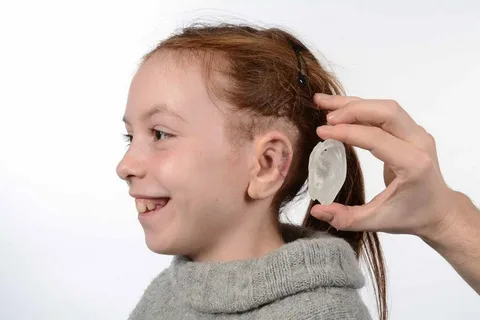
In a groundbreaking development, researchers from Weill Cornell Medicine and Cornell Engineering have utilized advanced tissue engineering methods along with 3D printing technology to create a replica of an adult human ear that closely mimics the look and feel of a natural ear. Published online in Acta Biomaterialia on March 16, the study showcases the potential of producing grafts with precise anatomical details and the appropriate biomechanical properties for individuals born with a congenital ear malformation or who have lost an ear later in life.
Ear reconstruction typically involves multiple surgeries and intricate artistic skills, as stated by Dr. Jason Spector, the chief of the Division of Plastic and Reconstructive Surgery at NewYork-Presbyterian/Weill Cornell Medical Center and a professor at Weill Cornell Medicine. This innovative technology could potentially offer a realistic option for thousands in need of surgery to correct outer ear deformities.
Traditionally, surgeons have reconstructed ears using cartilage harvested from a child’s ribs, a procedure that can be painful and result in scarring. While the resulting graft may resemble the recipient’s other ear, it lacks the same flexibility. Employing chondrocytes, the cells responsible for cartilage formation, could lead to a more natural-looking replacement ear. Previous attempts using animal-derived chondrocytes within a collagen scaffold showed initial success but lacked the ear’s distinct features due to contraction over time.
To tackle this challenge, Dr. Spector and his team utilized sterilized, animal-derived cartilage devoid of immune-triggering elements. This cartilage was incorporated into detailed, ear-shaped plastic scaffolds produced via a 3D printer using ear data from an individual. The cartilage fragments serve as internal reinforcement, fostering new tissue growth within the scaffold to strengthen the graft and prevent contraction, akin to rebar in construction.
In the span of three to six months, the structure evolved into cartilage-containing tissue that closely mirrored the ear’s anatomical structures, including the helical rim, anti-helix rim-inside-the-rim, and central, conchal bowl. This achievement marks a significant advancement in ear reconstruction, noted Dr. Spector.
Biomechanical assessments conducted with Dr. Larry Bonassar, a biomedical engineering expert at Cornell University, confirmed that the replicas exhibited flexibility and elasticity similar to human ear cartilage, although the engineered material was slightly less robust and prone to tearing. To enhance strength and durability, Dr. Spector intends to introduce chondrocytes derived from a minor cartilage sample from the recipient’s remaining ear. This approach aims to bolster the elastic proteins crucial for ear cartilage resilience, resulting in a graft more biomechanically akin to the natural ear.
*Note:
1. Source: Coherent Market Insights, Public sources, Desk research
2. We have leveraged AI tools to mine information and compile it


