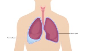
The pleura are two thin layers of tissue that line the lungs and chest cavity. The inner layer called the visceral pleura covers the outside of each lung. The outer layer called the parietal pleura lines the inner chest wall and diaphragm. Between these two layers is a small space called the pleural space that contains a thin film of fluid that allows the lungs to move smoothly within the chest cavity during breathing.
Pleural Effusion
Pleural effusion refers to an abnormal accumulation of excess fluid in the pleural space between the two layers of pleura. This buildup of fluid results in compression of the lungs which can make breathing difficult. Some common causes of a pleural effusion include:
– Congestive heart failure – When the heart is not pumping effectively, blood returning to the heart from the lungs backs up and fluid leaks into the chest cavity and pleural space.
– Cirrhosis of the liver – Advanced scarring of the liver can cause a backup of blood and fluid congestion in the blood vessels of the lung, leaking into the pleural space.
– Pneumonia or pulmonary embolism – Infections or blood clots in the lungs can cause inflammation and leaking of fluid into the pleural space.
– Cancer – Certain cancers like lung cancer, breast cancer or lymphomas can spread (metastasize) to the lung and pleura, causing a pleural effusion.
– Kidney failure – When the kidneys are not properly removing waste and excess fluid from the blood, fluid backs up in the lungs.
Diagnosis usually involves a physical exam, chest x-ray and ultrasound of the lungs. A sample of the pleural fluid is also often analyzed in a lab to determine the cause. Treatment depends on the underlying cause but may involve draining excess fluid, prescribing diuretics or treating the primary disease.
Pleurisy
Pleurisy, also called pleuritis, is inflammation of the pleura usually caused by a viral or bacterial infection. It results in chest pain that becomes worse with deep breathing, coughing or sneezing. Symptoms include sharp, stabbing pain in the chest wall, shortness of breath, fever and cough with sputum production.
The most common infectious causes are viruses like influenza, adenovirus or SARS-CoV-2. Bacterial pathogens like streptococcus or mycoplasma can also lead to pleurisy. Injury or conditions like pulmonary embolism, rheumatoid arthritis or lupus may also trigger pleural inflammation.
Diagnosis is based on symptoms, physical exam and chest x-ray findings. Blood tests may help rule out other potential causes of chest pain. Most cases resolve on their own with rest and over-the-counter pain medications. Antibiotics may be prescribed if a bacterial infection is suspected. Repeated bouts of pleurisy could indicate an underlying condition requiring additional treatment or management.
Pneumothorax
A pneumothorax occurs when air leaks into the pleural space between the lung and chest wall, causing the lung to collapse. It is a medical emergency that requires prompt treatment. There are two main types – spontaneous and traumatic:
– Spontaneous pneumothorax happens for no apparent reason, often affecting tall, thin young males. It occurs when a small tear forms in the pleura lining the lung.
– Traumatic pneumothorax results from a chest injury or medical procedure that punctures the lung, allowing air leak. It commonly follows injuries from car accidents, stab/gunshot wounds or ventilator use.
Symptoms include acute chest pain that worsens with breathing, shortness of breath, rapid breathing and heart rate. Diagnosis is by chest x-ray showing the collapsed lung. Treatment ranges from simple observation to chest tube insertion to reinflate the lung and allow healing. Surgery may be required for recurrent or persistent pneumothoraces.
Malignant Pleural Mesothelioma
Malignant pleural mesothelioma is a rare but aggressive form of cancer that develops in the mesothelial cells of the pleura. Significant risk factors include prolonged occupational or accidental exposure to asbestos fibers. Other possible causes are radiation, simian virus 40 and genetic factors.
Early symptoms are nonspecific like chest wall pain, breathlessness, persistent cough or weight loss. As the diseaseprogresses, signs include large pleural effusions, pleural thickening and respiratory failure. Diagnosis relies on pleural fluid analysis, imaging tests and thoracoscopic biopsy of the pleura.
Staging classifies the extent of disease spread. Prognosis tends to be poor with a median survival of 9-12 months. Treatment aims at palliation and may combine surgery, chemotherapy and radiotherapy depending on tumor stage. However, mesothelioma remains very difficult to treat effectively especially in cases of metastatic spread. More research continues on improving available therapies.
*Note:
1. Source: Coherent Market Insights, Public sources, Desk research
2. We have leveraged AI tools to mine information and compile it


